Keratinocyte
หน้าตา
บทความนี้มีชื่อเป็นภาษาอังกฤษ เนื่องจากยังไม่มีชื่อภาษาไทยที่กระชับ เหมาะสม, ไม่ปรากฏคำอ่านที่แน่ชัด หรือไม่ปรากฏคำแปลที่ใช้ในทางวิชาการ |


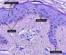
Keratinocyte เป็นเซลล์ประเภทหลักที่พบในหนังกำพร้าซึ่งเป็นผิวหนังชั้นนอกสุด ผิวหนังชั้นนอกสุดของมนุษย์ 90% เป็นเซลล์ชนิดนี้[1] มีหน้าที่ป้องกันผิวหนังจากอันตรายเนื่องกับสิ่งแวดล้อมรวมทั้งความร้อน รังสีอัลตราไวโอเลต การสูญเสียน้ำ และจุลชีพก่อโรคต่าง ๆ เช่น แบคทีเรีย เชื้อรา ปรสิต และไวรัส มีโครงสร้างที่เป็นโปรตีน เอนไซม์ ลิพิด และเพปไทด์ต้านจุลชีพ (antimicrobial peptide) ที่อำนวยหน้าที่ป้องกันอันตรายเช่นนี้ของผิวหนัง เซลล์เปลี่ยนสภาพเริ่มต้นจากเซลล์ต้นกำเนิดหนังกำพร้า (epidermal stem cells) ในส่วนล่างของหนังกำพร้า แล้วย้ายขึ้นไปสู่ผิวหนัง โดยสุดท้ายกลายเป็น corneocyte แล้วในที่สุดก็จะลอกออกจากผิวหนัง[2][3][4][5] โดยในมนุษย์เกิดขึ้นทุก ๆ 40-56 วัน[6] basal cell ในผิวหนังชั้นฐาน (stratum basale) บางครั้งเรียกว่า basal keratinocytes[7]
ดูเพิ่ม
[แก้]เชิงอรรถและอ้างอิง
[แก้]- ↑ McGrath JA; Eady RAJ; Pope FM. (2004). "Anatomy and Organization of Human Skin". ใน Burns T; Breathnach S; Cox N; Griffiths C. (บ.ก.). Rook's Textbook of Dermatology (7th ed.). Blackwell Publishing. p. 4190. doi:10.1002/9780470750520.ch3. ISBN 978-0-632-06429-8. คลังข้อมูลเก่าเก็บจากแหล่งเดิมเมื่อ 2020-05-20. สืบค้นเมื่อ 2010-06-01.
- ↑
Gilbert, Scott F. (2000). "The Epidermis and the Origin of Cutaneous Structures.". Developmental Biology. Sinauer Associates. ISBN 978-0878932436.
Throughout life, the dead keratinized cells of the cornified layer are shed (humans lose about 1.5 grams of these cells each day*) and are replaced by new cells, the source of which is the mitotic cells of the Malpighian layer. Pigment cells (melanocytes) from the neural crest also reside in the Malpighian layer, where they transfer their pigment sacs (melanosomes) to the developing keratinocytes.
- ↑ Houben, E; De Paepe, K; Rogiers, V (2007). "A keratinocyte's course of life". Skin Pharmacology and Physiology. 20 (3): 122–32. doi:10.1159/000098163. PMID 17191035. S2CID 25671082.
- ↑ Ghazizadeh, S; Taichman, LB (March 2001). "Multiple classes of stem cells in cutaneous epithelium: a lineage analysis of adult mouse skin". The EMBO Journal. 20 (6): 1215–22. doi:10.1093/emboj/20.6.1215. PMC 145528. PMID 11250888.
- ↑ Koster MI (July 2009). "Making an epidermis". Annals of the New York Academy of Sciences. 1170 (1): 7–10. Bibcode:2009NYASA1170....7K. doi:10.1111/j.1749-6632.2009.04363.x. PMC 2861991. PMID 19686098.
- ↑ Halprin KM (January 1972). "Epidermal "turnover time"--a re-examination". The British Journal of Dermatology. 86 (1): 14–9. doi:10.1111/j.1365-2133.1972.tb01886.x. PMID 4551262. S2CID 30165907.
- ↑ James, W; Berger, T; Elston, D (December 2005). Andrews' Diseases of the Skin: Clinical Dermatology (10th ed.). Saunders. pp. 5–6. ISBN 978-0-7216-2921-6. คลังข้อมูลเก่าเก็บจากแหล่งเดิมเมื่อ 2010-10-11. สืบค้นเมื่อ 2010-06-01.
แหล่งข้อมูลอื่น
[แก้]วิกิมีเดียคอมมอนส์มีสื่อที่เกี่ยวข้องกับ Keratinocyte
- Tang, L.; Wu, J.J.; Ma, Q.; Cui, T.; Andreopoulos, F.M.; Gil, J.; Valdes, J.; Davis, S.C.; Li, J. (2010-03-06). "Human lactoferrin stimulates skin keratinocyte function and wound re-epithelialization". British Journal of Dermatology. Wiley. 163 (1): 38–47. doi:10.1111/j.1365-2133.2010.09748.x. ISSN 0007-0963.
