
ไฟล์:Cajal Retina.jpg
ดูภาพที่มีความละเอียดสูงกว่า (500 × 745 พิกเซล, ขนาดไฟล์: 83 กิโลไบต์, ชนิดไมม์: image/jpeg)
| รูปภาพหรือไฟล์เสียงนี้ ต้นฉบับอยู่ที่ คอมมอนส์ รายละเอียดด้านล่าง เป็นข้อความที่แสดงผลจาก ไฟล์ต้นฉบับในคอมมอนส์
|
ความย่อ
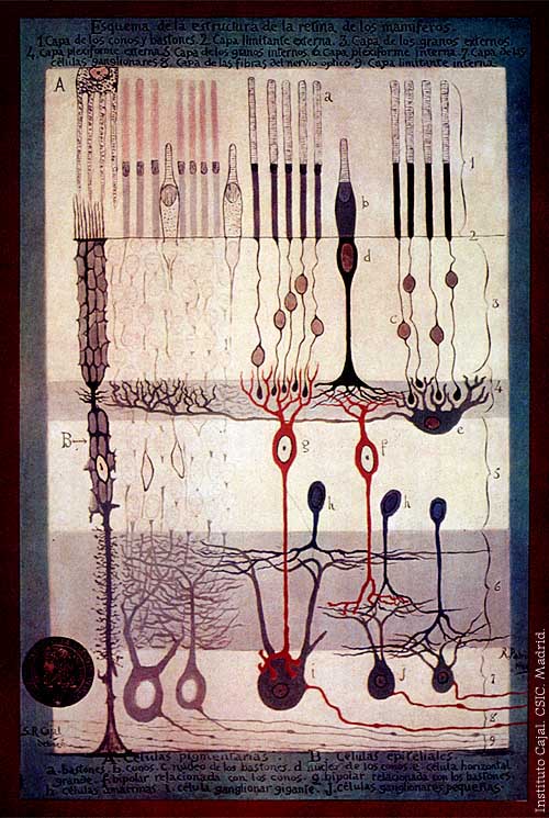
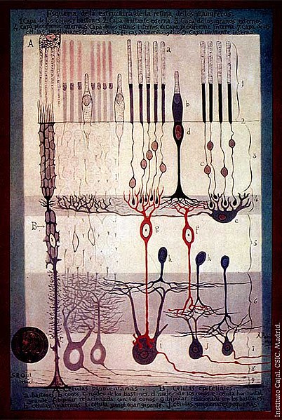
From "Structure of the Mammalian Retina" c.1900 By Santiago Ramon y Cajal.
Outline of the structure of the mammalian retina. 1. Rod and cone layer. 2. External limiting membrane. 3. Outer granular layer. 4. Outer plexiform layer. 5. Inner granular layer. 6. Inner plexiform layer. 7. Ganglion cell layer. 8. Optic nerve fibre layer. 9. Internal limiting membrane. A. Pigmented cells. B. Epithelial cells. a. Rods. b. Cones. c. Rod nucleus. d. Cone Nucleus. e. Large horizontal cell f. Cone-associated bipolar cell. g. Rod-associated bipolar cell. h. Amacrine cells. i. Giant ganglion cell. j. Small ganglion cells.
การอนุญาตใช้สิทธิ
|
งานนี้เป็นสาธารณสมบัติ ในประเทศต้นกำเนิดและประเทศอื่น ๆ ที่ระยะเวลาการคุ้มครองลิขสิทธิ์น้อยกว่า 70 ปีหลังจากผู้สร้างสรรค์งานเสียชีวิต.
| |
| ไฟล์นี้ได้ถูกระบุว่าไม่มีข้อจำกัดภายใต้กฎหมายลิขสิทธิ์ รวมถึงสิทธิที่เกี่ยวข้องและที่ใกล้เคียงกัน | |
ประวัติไฟล์
คลิกวันที่/เวลาเพื่อดูไฟล์ที่ปรากฏในขณะนั้น
| วันที่/เวลา | รูปย่อ | ขนาด | ผู้ใช้ | ความเห็น | |
|---|---|---|---|---|---|
| ปัจจุบัน | 00:42, 5 มีนาคม 2549 | | 500 × 745 (83 กิโลไบต์) | Feezil~commonswiki | From "Structure of the Mammalian Retina" c.1900 By Santiago Ramon y Cajal. 1.- Rod and Cone layer 2.-Outer nuclear layer 3.- Granule layer 4.- External plexiform layer A: Pigmented cells; B: epithelial cells |
หน้าที่มีภาพนี้
หน้าต่อไปนี้ โยงมาที่ภาพนี้:
การใช้ไฟล์ข้ามโครงการ
วิกิอื่นต่อไปนี้ใช้ไฟล์นี้:
- การใช้บน ar.wikipedia.org
- การใช้บน bn.wikipedia.org
- การใช้บน ca.wikipedia.org
- การใช้บน en.wikipedia.org
- การใช้บน en.wikiversity.org
- Human vision and function/Part 1: How the eye works/1.3 Light stimulus and the eye
- User:Jtwsaddress42/People/Ramón y Cajal, Santiago
- User:Jtwsaddress42/People/R
- User:Jtwsaddress42/Gallery/Ramón y Cajal, Santiago
- User:Jtwsaddress42/Gallery/Ramón y Cajal, Santiago - The Visual System
- User:Jtwsaddress42/Gallery
- การใช้บน es.wikipedia.org
- การใช้บน et.wikipedia.org
- การใช้บน ext.wikipedia.org
- การใช้บน fa.wikipedia.org
- การใช้บน fr.wikipedia.org
- การใช้บน gl.wikipedia.org
- การใช้บน he.wikipedia.org
- การใช้บน hy.wikipedia.org
- การใช้บน it.wikipedia.org
- การใช้บน ja.wikipedia.org
- การใช้บน ko.wikipedia.org
- การใช้บน ml.wikipedia.org
- การใช้บน pt.wikipedia.org
- การใช้บน ru.wikipedia.org
- การใช้บน simple.wikipedia.org
- การใช้บน uk.wikipedia.org
- การใช้บน vi.wikipedia.org
- การใช้บน zh.wikipedia.org