
ไฟล์:Rabies encephalitis Negri bodies PHIL 3377 lores.jpg

ขนาดของตัวอย่างนี้: 800 × 532 พิกเซล ความละเอียดอื่น: 320 × 213 พิกเซล | 640 × 425 พิกเซล | 1,024 × 681 พิกเซล | 1,280 × 851 พิกเซล | 1,801 × 1,197 พิกเซล
ดูภาพที่มีความละเอียดสูงกว่า (1,801 × 1,197 พิกเซล, ขนาดไฟล์: 438 กิโลไบต์, ชนิดไมม์: image/jpeg)
| รูปภาพหรือไฟล์เสียงนี้ ต้นฉบับอยู่ที่ คอมมอนส์ รายละเอียดด้านล่าง เป็นข้อความที่แสดงผลจาก ไฟล์ต้นฉบับในคอมมอนส์
|
ความย่อ
| คำอธิบาย |
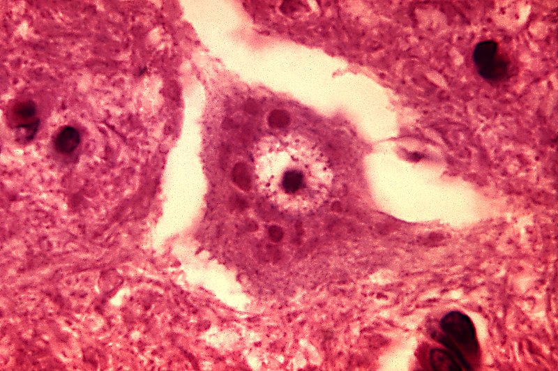
English: This micrograph depicts the histopathologic changes associated with rabies encephalitis prepared using an H&E stain.
Note the Negri bodies, which are cellular inclusions found most frequently in the pyramidal cells of Ammon's horn, and the Purkinje cells of the cerebellum. They are also found in the cells of the medulla and various other ganglia. |
||
| วันที่ | |||
| แหล่งที่มา |
|
||
| ผู้สร้างสรรค์ | Content Provider(s): CDC/Dr. Daniel P. Perl | ||
| การอนุญาต (การใช้ไฟล์นี้ใหม่) |
Copyright Restrictions: None – This image is in the public domain and thus free of any copyright restrictions. As a matter of courtesy we request that the content provider be credited and notified in any public or private usage of this image. | ||
| เวอร์ชันอื่น |
 |
การอนุญาตใช้สิทธิ
This image is a work of the Centers for Disease Control and Prevention, part of the United States Department of Health and Human Services, taken or made as part of an employee's official duties. As a work of the U.S. federal government, the image is in the public domain.
|
ประวัติไฟล์
คลิกวันที่/เวลาเพื่อดูไฟล์ที่ปรากฏในขณะนั้น
| วันที่/เวลา | รูปย่อ | ขนาด | ผู้ใช้ | ความเห็น | |
|---|---|---|---|---|---|
| ปัจจุบัน | 14:27, 26 ตุลาคม 2554 | | 1,801 × 1,197 (438 กิโลไบต์) | Ghainmem | Higher-resolution version |
| 01:01, 31 พฤษภาคม 2549 |  | 700 × 465 (57 กิโลไบต์) | Patho | {{Information| |Description=ID#: 3377 Description: This micrograph depicts the histopathologic changes associated with rabies encephalitis prepared using an H&E stain. Note the Negri bodies, which are cellular inclusions found most frequently in the pyra |
หน้าที่มีภาพนี้
หน้าต่อไปนี้ โยงมาที่ภาพนี้:
การใช้ไฟล์ข้ามโครงการ
วิกิอื่นต่อไปนี้ใช้ไฟล์นี้:
- การใช้บน ar.wikipedia.org
- การใช้บน de.wikibooks.org
- การใช้บน fr.wikipedia.org
- การใช้บน ja.wikipedia.org
- การใช้บน uk.wikipedia.org