
ไฟล์:PBP catalysis.svg

ดูภาพที่มีความละเอียดสูงกว่า ((ไฟล์ SVG, 1,142 × 1,567 พิกเซล, ขนาดไฟล์: 1.44 เมกะไบต์))
| รูปภาพหรือไฟล์เสียงนี้ ต้นฉบับอยู่ที่ คอมมอนส์ รายละเอียดด้านล่าง เป็นข้อความที่แสดงผลจาก ไฟล์ต้นฉบับในคอมมอนส์
|
ความย่อ
| คำอธิบาย |
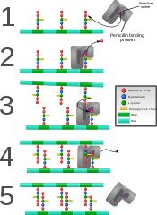
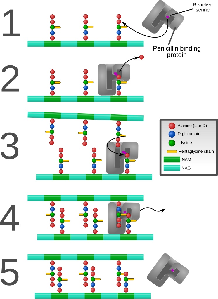
English: Diagram depicting formation of cross-links in the bacterial cell wall by a penicillin binding protein (PBP, an enzyme).
1. The bacterial cell wall consists of strands of repeating N-acetylglucosamine (NAG) and N-acetylmuramic acid (NAM) subunits. The NAM subunits have short peptide chains attached to them. (The exact composition of these can vary. The proximal alanine is usually L-ala and the distal two are usually D-ala.) These chains, in turn, are bound to chains of 5 glycine residues that will be used in cross-linking. 2. The PBP forms a bond with the peptide side chain at the second most distal alanine residue. This displaces the most distal alanine residue. 3. Another strand of bacterial cell wall arrives. The free end of one of the pentaglycine chains displaces the PBP and forms a bond with the terminal alanine on the other strand. 4. After being displaced, the PBP diffuses away. 5. The formation of one cross-link is complete. |
| วันที่ | |
| แหล่งที่มา | งานของตัว |
| ผู้สร้างสรรค์ | Mcstrother |
| เวอร์ชันอื่น |
[]
|


การอนุญาตใช้สิทธิ
- คุณสามารถ:
- ที่จะแบ่งปัน – ที่จะทำสำเนา แจกจ่าย และส่งงานดังกล่าวต่อไป
- ที่จะเรียบเรียงใหม่ – ที่จะดัดแปลงงานดังกล่าว
- ภายใต้เงื่อนไขต่อไปนี้:
- แสดงที่มา – คุณต้องให้เกียรติเจ้าของงานอย่างเหมาะสม โดยเพิ่มลิงก์ไปยังสัญญาอนุญาต และระบุหากมีการเปลี่ยนแปลง คุณอาจทำเช่นนี้ได้ในรูปแบบใดก็ได้ตามควร แต่ต้องไม่ใช่ในลักษณะที่แนะว่าผู้ให้อนุญาตสนับสนุนคุณหรือการใช้งานของคุณ
ประวัติไฟล์
คลิกวันที่/เวลาเพื่อดูไฟล์ที่ปรากฏในขณะนั้น
| วันที่/เวลา | รูปย่อ | ขนาด | ผู้ใช้ | ความเห็น | |
|---|---|---|---|---|---|
| ปัจจุบัน | 02:26, 10 กันยายน 2554 | | 1,142 × 1,567 (1.44 เมกะไบต์) | Mcstrother | Major revision. Corrected inaccuracies in previous image. |
| 11:15, 3 พฤษภาคม 2554 |  | 1,139 × 1,062 (850 กิโลไบต์) | Mcstrother | Changed all fonts to Liberation Sans | |
| 10:46, 10 เมษายน 2554 |  | 1,139 × 1,062 (850 กิโลไบต์) | Mcstrother | Changed color of carbohydrate chain. | |
| 10:30, 7 มีนาคม 2554 |  | 1,139 × 1,062 (835 กิโลไบต์) | Mcstrother | {{Information |Description ={{en|1=Diagram depicting formation of cross-links in the bacterial cell wall by a penicillin binding protein (PBP, an enzyme). 1. The bacterial cell wall consists of strands of repeating N-acetylglucosamine (NAG) and N-ace |
หน้าที่มีภาพนี้
หน้าต่อไปนี้ โยงมาที่ภาพนี้:
การใช้ไฟล์ข้ามโครงการ
วิกิอื่นต่อไปนี้ใช้ไฟล์นี้:
- การใช้บน en.wikipedia.org
- การใช้บน es.wikipedia.org
- การใช้บน fa.wikipedia.org
- การใช้บน ga.wikipedia.org
- การใช้บน gl.wikipedia.org
- การใช้บน hu.wikipedia.org
- การใช้บน it.wikipedia.org
- การใช้บน mk.wikipedia.org