
ไฟล์:Nematocyst discharge.png
ไม่มีภาพที่มีรายละเอียดสูงกว่านี้
Nematocyst_discharge.png (480 × 371 พิกเซล, ขนาดไฟล์: 190 กิโลไบต์, ชนิดไมม์: image/png)
| รูปภาพหรือไฟล์เสียงนี้ ต้นฉบับอยู่ที่ คอมมอนส์ รายละเอียดด้านล่าง เป็นข้อความที่แสดงผลจาก ไฟล์ต้นฉบับในคอมมอนส์
|

|
ภาพทางชีววิทยานี้ ควรจะถูกสร้างใหม่เป็นกราฟิกส์เวกเตอร์ ซึ่งมีข้อดีอีกหลายประการ ดูเพิ่มเติมที่คอมมอนส์:สื่อต้องการเก็บกวาดสำหรับสารสนเทศเพิ่มเติม หากภาพนี้มีในรูปแบบกราฟิกส์เวกเตอร์อยู่แล้ว กรุณาอัปโหลดและแทนที่แม่แบบนี้ด้วย
{{vector version available|ชื่อภาพใหม่}}
แนะนำเป็นอย่างยิ่งให้ตั้งชื่อไฟล์เวกเตอร์ใหม่นั้นในรูปแบบ "Nematocyst discharge.svg" แล้วใส่แม่แบบ Vector version available (หรือ Vva) ซึ่งไม่ต้องใช้พารามิเตอร์ ชื่อภาพใหม่ |
ความย่อ
| คำอธิบาย |
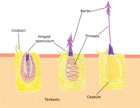
English: The diagram above shows the anatomy of a nematocyst cell and its “firing” sequence, from left to right. On the far left is a nematocyst inside its cellular capsule. The cell’s thread is coiled under pressure and wrapped around a stinging barb. When potential prey makes contact with the tentacles of a polyp, the nematocyst cell is stimulated. This causes a flap of tissue covering the nematocyst—the operculum—to fly open. The middle image shows the open operculum, the rapidly uncoiling thread and the emerging barb. On the far right is the fully extended cell. The barbs at the end of the nematocyst are designed to stick into the polyp’s victim and inject a poisonous liquid. When subdued, the polyp’s tentacles move the prey toward its mouth and the nematocysts recoil back into their capsules. |
| วันที่ | 11 เมษายน พ.ศ. 2550 (original upload date) |
| แหล่งที่มา | นำเข้าจาก en.wikipedia มายังคอมมอนส์ |
| ผู้สร้างสรรค์ | The original uploader was Spaully ที่ วิกิพีเดียภาษาอังกฤษ. |
การอนุญาตใช้สิทธิ
This file is licensed under Creative Commons ShareAlike 1.0 License.
Creative Commons has retired this legal tool and does not recommend that it be applied to works.
|
This image is in the public domain because it contains materials that originally came from the U.S. National Oceanic and Atmospheric Administration, taken or made as part of an employee's official duties.
|
บันทึกการอัพโหลด
The original description page was here. All following user names refer to en.wikipedia.
- 2007-04-11 17:10 Spaully 480×371×8 (194868 bytes) Modified from: http://www.oceanservice.noaa.gov/education/kits/corals/media/supp_coral01b.html {{Information |Description=Nematocyst discharge process. |Source= Modified from [http://www.oceanservice.noaa.gov/education/kits/corals/media/supp_coral01b.html
ประวัติไฟล์
คลิกวันที่/เวลาเพื่อดูไฟล์ที่ปรากฏในขณะนั้น
| วันที่/เวลา | รูปย่อ | ขนาด | ผู้ใช้ | ความเห็น | |
|---|---|---|---|---|---|
| ปัจจุบัน | 00:29, 14 ตุลาคม 2550 | | 480 × 371 (190 กิโลไบต์) | Alison | {{Information |Description===Description== The diagram above shows the anatomy of a nematocyst cell and its “firing” sequence, from left to right. On the far left is a nematocyst inside its cellular capsule. The cell’s thread is coiled under pressur |
หน้าที่มีภาพนี้
หน้าต่อไปนี้ โยงมาที่ภาพนี้:
การใช้ไฟล์ข้ามโครงการ
วิกิอื่นต่อไปนี้ใช้ไฟล์นี้:
- การใช้บน ca.wikipedia.org
- การใช้บน ceb.wikipedia.org
- การใช้บน en.wikipedia.org
- การใช้บน fr.wikipedia.org
- การใช้บน hr.wikipedia.org
- การใช้บน id.wikipedia.org
- การใช้บน it.wikibooks.org
- การใช้บน ja.wikipedia.org
- การใช้บน lv.wikipedia.org
- การใช้บน ms.wikipedia.org
- การใช้บน my.wikipedia.org
- การใช้บน pa.wikipedia.org
- การใช้บน pt.wikipedia.org
- การใช้บน simple.wikipedia.org
- การใช้บน sv.wikipedia.org
- การใช้บน te.wikipedia.org
- การใช้บน vi.wikipedia.org